
More Information
Submitted: December 07, 2023 | Approved: December 22, 2023 | Published: December 26, 2023
How to cite this article: Pío-Rendón JB, Andreu FJV, Palacios DC, Mesa VD. Osseous Choristoma with in a Dermolipoma: 17 Years Old Girl Case Report. Int J Clin Exp Ophthalmol. 2023; 7: 008-010.
DOI: 10.29328/journal.ijceo.1001053
Copyright Licence: © 2023 Pío-Rendón JB, et al. This is an open access article distributed under the Creative Commons Attribution License, which permits unrestricted use, distribution, and reproduction in any medium, provided the original work is properly cited.

Osseous Choristoma with in a Dermolipoma: 17 Years Old Girl Case Report
Julian Bermúdez Pío-Rendón, Francisco Javier Vicente Andreu, David Cerdán Palacios and Vanessa Díaz Mesa*
Hospital Arruzafa, Córdoba, Spain
*Address for Correspondence: Vanessa Díaz Mesa, Hospital Arruzafa, Córdoba, Spain, Email: [email protected]
Bone choristoma within a dermolipoma is a rare epibulbar tumor with a low prevalence. It is a benign tumor that does not usually cause discomfort or functional problems to patients who suffer from it. Its treatment is surgical and with an aesthetic purpose.
We report the case of a 17-year-old patient with a bone choristoma, a tooth, within a dermolipoma.
Epibulbar bone choristoma is a rare benign tumor that causes little discomfort to patients who suffer from it and is asymptomatic in most cases. Computed tomography (CT) is essential in its diagnosis and its treatment is surgical, but always due to aesthetic reasons.
Among the different types of epibulbar tumors described [1], we can find complex choristomatous tumors, defined as the rarest type of choristoma with more than two normal tissues growing in an abnormal location. Rarely, a bone choristoma is seen within a dermolipoma in the epibulbar region [2]. Histopathologically, this complex tumor is composed of adipose tissue and compact mature bone.
Treatment of this type of tumor is surgical, for aesthetic reasons, since these benign tumors do not usually cause functional problems to patients.
We report a case of complex choristoma located in the epibulbar region. The challenge to find one of these cases is added, together with the rarity that bone choristoma is a tooth.
This report adheres to the Declaration of Helsinki and HIPAA compliance.
In February 2013, a 7-year-old girl attended her first visit with a mass in the upper epibulbar region of her left eye. This mass was present at birth and until that moment. It had been controlled by pediatricians who were not ophthalmologists, without apparent changes, including its size. Her uncorrected visual acuity reached LogMAR 0 on both eyes (OU), and intraocular pressure was 16mmHg OU. OU presented papillary conjunctivitis and the rest of the examination in the right eye was completely normal. In the left eye, a pinkish, smooth-surfaced, oval, and raised epibulbar mass was observed in the superotemporal quadrant with a palpable hard mass inside (Figure 1).
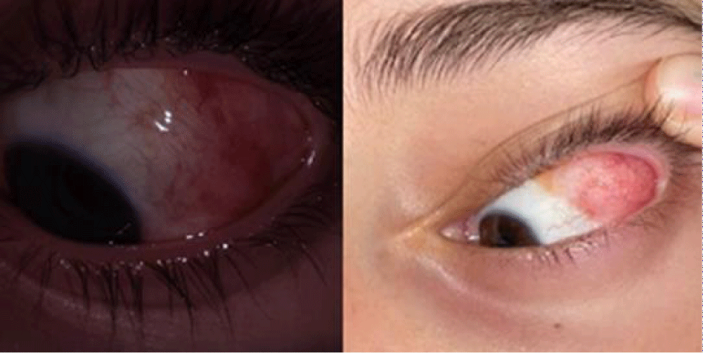
Figure 1: Clinical Presentation of the Left Eye in a 7-Year-Old Girl.
This lesion does not cause restrictions in eye movements, but it causes small palpebral ptosis and occasional discomfort. Artificial tears were prescribed and images were requested at the next annual check-up. A control visit was made in November of the same year, in which the patient provided orbital computed tomography (CT) images that revealed an oval cystic mass of low-density fat and a mass compatible with bone inside, corroborating the diagnosis of dermolipoma with bony choristoma. In this type of tumor, this imaging test is differential in diagnosis [3,4].
On successive controls, everything remained unchanged until April 2017, when the mild ptosis disappeared, maintaining the size and other characteristics of the tumor. Surgery was considered and the patient’s family preferred to wait. Annual controls followed and nothing changed, size and the rest of the characteristics of the cyst were controlled by photography to see that there was no evolution. In her control in April 2019, despite the fact that everything remained stable, the patient, who was already 13 years old, for the first time showed her willingness to undergo surgery, due to aesthetic discomfort, however, her guardians decided to wait. On the next control, in June 2022, the patient and her family, after verifying in the ophthalmological examination that everything remains the same, decided to accept the surgical solution. Excision of the dermolipoma under local anesthesia for cosmetic purposes was indicated.
The surgical technique begins with the preparation of the eye with a 2.5% phenylephrine eye drop as vasoconstrictor to improve the hemostasis of surgery, then a traction suture with a five-zero silk is used to retract the eye and mobilize it, to a most comfortable position to work on the tumor. After this, a subconjunctival lidocaine and bupivacaine anesthetic solution is used prior to tumor surgery. Next, conjunctiva is dissected, in order to obtain a complete dissection between conjunctiva and dermolipoma, trying to follow the same plane in all sections to achieve a dissection as clean as possible, preserving as much conjunctival tissue as possible to achieve a better posterior reconstruction of the same conjunctiva, both anatomical and aesthetic. However, in our case, the internal portion of the tumor is in contact with the lateral rectus muscle, making the dissection more complex, trying to preserve or maintain the structure and function of the muscle. In the upper portion of the dermolipoma, dissection must be very careful working near the lacrimal gland, trying not to damage it, in the excision of the tumor. Once the edges of the dermolipoma have been dissected, a posterior dissection was performed to extract it and send it to Pathological Anatomy. In our surgery, once all or a large part of the tumor had been dissected, we found an indurated, hard tissue with a solid appearance, adhered to the superior temporal conjunctiva of the eyeball. Tissue that we had to carefully dissect from the sclera, trying to avoid its tearing. Thus, when extracting this solid tissue (Figure 2), in the macroscopic examination, it corresponds to a mature bone tooth of an adult person. It was sent to the Pathological Anatomy for analysis, with histopathological confirmation. To finish the surgery, a reconstruction of the dissected conjunctiva was made using a reverse and discontinuous seven-zero vycril suture, trying to achieve the best aesthetic, as well as functional, result.
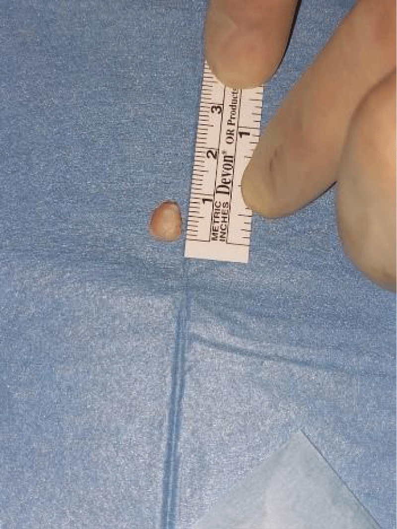
Figure 2: Surgical Procedure - Dissection of Dermolipoma and Solid Tissue.
Post-surgical treatment is corticosteroids and topical antibiotics in a descending pattern: we start with 5 drops/day for 5 days and gradually decrease to 1 drop/day for 5 days, along with a moisturizing treatment for the following weeks, until it is controlled.
On the first control carried out a week after surgery, the patient only complained of discomfort looking to the right side. On examination, everything is normal, except for a Corneal Dellen observed at 3 hours and conjunctival hyperemia (Figure 3).
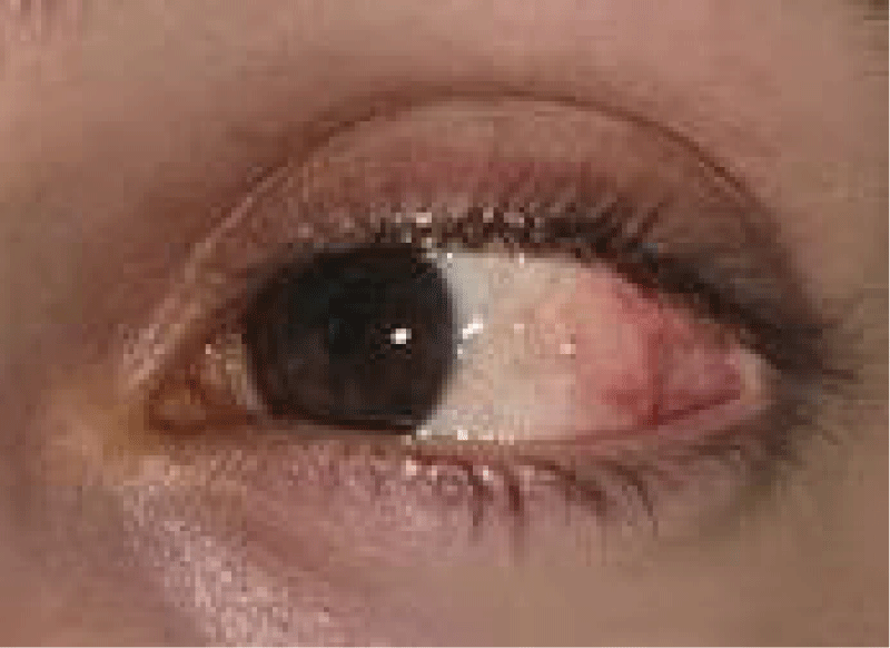
Figure 3: Post-Surgical Evaluation - Corneal Dellen and Conjunctival Hyperemia.
The patient was visited again, two weeks after surgery, she is better and on examination: Dellen has partially resolved, although it can still be observed and the hyperemia has improved, although it is still present. Treatment is not modified. A month and a half after surgery, both Dellen and hyperemia continue being observed, along with perfect healing. Three months after surgery, the patient is checked again, discovering that Dellen has disappeared and there is only a slight hyperemia over the conjunctival scar. Nine months after surgery, in the last control carried out, the patient reported irritation and discomfort in the left eye, especially when she was nervous. On examination, hyperemia was observed in the old dermolipoma area with some relief, a conjunctival cyst, which is pending evaluation in successive visits.
Bone choristoma, which corresponds to a tooth, within a dermolipoma in the epibulbar region is a rare tumor. In a 1975 study, by Elsas, et al. 302 epibulbar tumors in children were studied and only 6 had a diagnosis of complex Choristoma. This study also reported the presence of an immature tooth within a dermolipoma [1]. In the study by Cunha, et al, of 282 epibulbar tumors, only 15 dermolipomas were reported and within these, only one was reported as a bone choristoma [3,5].
As in our case, this type of tumor shows a female preponderance, it is not associated with systemic diseases [2] it does not usually cause discomfort to patients, and does not alter the functionality or anatomy of the eye or its annexes. Follow-up of the case is usual, although patients, as in our case, usually demand its excision for aesthetic reasons. The complexity of the surgery is to preserve the rest of the structures such as the lacrimal gland, extraocular muscles, or sclera itself.
Finding a bone choristoma within a dermo lipoma is very uncommon, especially if the bone tissue is a tooth. Computed tomography (CT) is an essential and differential test in its diagnosis and monitoring its evolution is the most common option. It is a tumor that usually does not bother patients and its excision, which must be very careful due to the proximity of other major structures, is basically performed for cosmetic reasons.
Patient consent
The patient’s family provided consent to publish details of this case.
Précis: Case report of an uncommon type of orbital tumor, Osseous choristoma within a dermolipoma: diagnostic and surgical treatment.
This report adheres to the Declaration of Helsinki and HIPAA compliance.
- Elsas FJ, Green WR. Epibulbar tumors in childhood. Am J Ophthalmol. 1975 Jun;79(6):1001-7. doi: 10.1016/0002-9394(75)90685-6. PMID: 166560.
- Herdiana TR, Takahashi Y, Valencia MRP, Ana-Magadia MG, Ishikawa E, Kakizaki H. Epibulbar osseous choristoma within a dermolipoma: case report and literature review. Orbit. 2019 Oct;38(5):407-411. doi: 10.1080/01676830.2018.1539110. Epub 2018 Nov 15. PMID: 30430897.
- Gayre GS, Proia AD, Dutton JJ. Epibulbar osseous choristoma: case report and review of the literature. Ophthalmic Surg Lasers. 2002 Sep-Oct;33(5):410-5. PMID: 12358295.
- Kim E, Kim HJ, Kim YD, Woo KI, Lee H, Kim ST. Subconjunctival fat prolapse and dermolipoma of the orbit: differentiation on CT and MR imaging. AJNR Am J Neuroradiol. 2011 Mar;32(3):465-7. doi: 10.3174/ajnr.A2313. Epub 2010 Dec 16. PMID: 21163882; PMCID: PMC8013083.
- Cunha RP, Cunha MC, Shields JA. Epibulbar tumors in children: a survey of 282 biopsies. J Pediatr Ophthalmol Strabismus. 1987 Sep-Oct;24(5):249-54. doi: 10.3928/0191-3913-19870901-13. PMID: 3681613.