
More Information
Submitted: August 19, 2026 | Accepted: August 22, 2026 | Published: August 24, 2026
Citation: Chintamani P, Singh A, Menda PN, Keni NA, Gawde SS. To Compare the Accuracy of the Borish Delayed Technique and Cycloplegic Refraction in Determining Refractive Errors among Children Aged 6–18 Years. Int J Clin Exp Ophthalmol. 2026; 10(2): 6-9. Available from:
https://dx.doi.org/10.29328/journal.ijceo.1001065.
DOI: 10.29328/journal.ijceo.1001065
Copyright Licence: © 2026 Chintamani P, et al. This is an open access article distributed under the Creative Commons Attribution License, which permits unrestricted use, distribution, and reproduction in any medium, provided the original work is properly cited.
Keywords: Cycloplegic refraction; Borish delayed refraction; Pediatric refraction; Latent hyperopia; Control of accommodation; Assessment of the refractive error; Child myopia

To Compare the Accuracy of the Borish Delayed Technique and Cycloplegic Refraction in Determining Refractive Errors among Children Aged 6–18 Years
Prakash Chintamani, Anju Singh*, Purushottam Naidu Menda, Niyati Arun Keni and Swanandi Shriram Gawde
Department of Optometry, ITM Institute of Health Sciences, ITM Skills University, Navi Mumbai, Maharashtra, India
*Address for Correspondence: Anju Singh, Department of Optometry, ITM Institute of Health Sciences, ITM Skills University, Navi Mumbai, Maharashtra, India, Email: [email protected]
Aim: To compare spherical equivalent refractive measurements obtained using the Borish delayed technique and cycloplegic refraction in children and to assess the agreement between the two techniques.
Settings and design: Comparative Cross-Sectional Study.
Methods and materials: A comparative cross-sectional study was done among 51 children who were 7-16 years of age. Each participant underwent comprehensive ocular examination followed by dry refraction, Borish delayed refraction, and cycloplegic refraction. Spherical equivalent values obtained by the Borish delayed technique and cycloplegic refraction were compared for both eyes. Normality of the data was assessed using the Shapiro–Wilk test. As the data were nonnormally distributed, non-parametric statistical analyses were performed. The Wilcoxon signedrank test was used to compare paired measurements, the Mann–Whitney U test was used for gender-wise comparisons, and Spearman’s rank correlation coefficient was used to evaluate associations.
Results: 51 participants were included in the study. The mean SE refractive power revealed a progressive unmasking of latent hyperopia across techniques with Cycloplegic Refraction: 1.53 ± 3.08 (RE) and 1.36 ± 2.79 (LE) and Borish delayed technique: 1.01 ± 3.03 (RE) and 1.03 ± 2.72 (LE) Wilcoxon Signed-Rank tests showed a statistically significant difference between Cycloplegic Refraction and the Borish Delayed Technique for both the Right Eye (W = 1172, p < 0.05, Effect Size = 0.993) and Left Eye (W = 978, p < 0.05, Effect Size = 0.809). Cycloplegic refraction revealed a mean additional latent hyperopia of +0.52 D in RE and +0.33 D in LE.
Conclusion: The Borish delayed technique may be considered an adjunctive approach when cycloplegic refraction is not feasible, but it should not be regarded as interchangeable with cycloplegic refraction on the basis of statistical difference testing alone. Nevertheless, cycloplegic refraction remains the gold standard for accurate assessment of pediatric refractive errors, particularly for detecting latent hyperopia.
Refractive errors remain one of the leading causes of visual impairment among children worldwide and constitute a significant public health concern. Accurate assessment of refractive status during childhood is essential for normal visual development, prevention of amblyopia, and timely correction of accommodative and binocular vision disorders. Failure to detect refractive errors accurately may result in reduced academic performance, visual discomfort, and long-term visual consequences [1–4].
Accommodation presents a major challenge during pediatric refraction. Active accommodation frequently masks latent hyperopia, resulting in underestimation of hyperopic refractive errors during non-cycloplegic examinations [5,6]. Cycloplegic refraction temporarily eliminates accommodation through pharmacological cycloplegia and is therefore regarded as the gold standard for pediatric refractive assessment [7–9]. Despite its superior accuracy, cycloplegic refraction requires additional clinical time, pharmacological agents, patient cooperation, and post-examination recovery [8,9].
Several non-cycloplegic techniques have been developed to reduce accommodative influence while avoiding the disadvantages associated with cycloplegia. One such technique is the Borish delayed method, which attempts to relax accommodation before subjective refraction by incorporating a delay period and appropriate fogging procedures [10,11]. Previous studies have suggested that the Borish delayed technique may provide refractive values closer to cycloplegic findings than conventional dry refraction [11–13]. However, evidence comparing these techniques remains limited, particularly among Indian pediatric populations [13,14].
The present study therefore aimed to compare cycloplegic refraction, and the Borish delayed technique in determining accurate refractive errors in children and to evaluate whether the Borish delayed technique can serve as a clinically acceptable alternative when cycloplegic refraction is impractical.
Study design and setting
This prospective, comparative, cross-sectional study was conducted in the Department of Optometry at ITM Skills University Campus, Navi Mumbai, India for over a period of two years (2023–2025) to compare the accuracy of the Borish delayed technique with cycloplegic refraction in determining refractive errors among children.
The study was approved by the Institutional Ethics Committee of ITM IHS College of Nursing, Navi Mumbai, India (Approval No. ITMIHSCON/220925/3473, dated 22nd September 2025).
Written informed consent was obtained from the parent/legal guardian of each participant. Assent was obtained from children aged 7 above who were developmentally capable of providing assent.
Sample size
A formal a priori sample-size calculation was not performed. Participants were recruited using convenience sampling based on the availability of eligible participants during the study period, and 51 participants were included in the final analysis.
Sampling technique: A total of 51 participants aged 7–16 years with refractive errors were recruited using nonprobability convenience sampling by approaching the participants who turned up at the clinic during the study period.
Analyst blinding: The statistical analysis was performed using coded data; however, the analyst was not blinded to the measurement technique.
Children with normal ocular health who were cooperative for refraction were included in the study, while those with manifest strabismus, ocular pathology, previous ocular surgery, systemic diseases affecting accommodation or vision, allergy to cycloplegic agents, or poor cooperation were excluded.
All 51 participants had complete refractive measurements for both techniques, and no missing data were present for the variables included in the primary analysis.
Refraction procedures
Borish delayed subjective refraction: Objective refraction was first obtained using an autorefractometer followed by streak retinoscopy. Subjective refraction was subsequently performed according to the Borish delayed technique. To minimize accommodative influence, participants were instructed to maintain fixation on a distant target for approximately 10–15 minutes before the final refinement.
Cycloplegic refraction: Cycloplegic refraction was performed by using the drug cyclopentolate hydrochloride 1% with the help of streak retinoscope. Two drops were instilled into each eye at an interval of five minutes. Refraction was performed 30–45 minutes after the final instillation, once adequate cycloplegia had been confirmed by pupillary dilatation and a markedly reduced pupillary light reflex. The final refractive findings were spherical, cylindrical, axis, spherical equivalent and best-corrected visual acuity recorded
Statistical analysis
Data were analysed using Jamovi (Version 2.6). Continuous variables were expressed as mean ± standard deviation (SD), median, and interquartile range (IQR), while categorical variables were presented as frequencies and percentages. Normality was assessed using the Shapiro–Wilk test. Since the data were not normally distributed, the Wilcoxon signed-rank test was used to compare the Borish delayed technique and cycloplegic refraction, the Mann–Whitney U test was used for gender comparisons, and Spearman’s rank correlation was used to assess associations between variables. A p - value < 0.05 was considered statistically significant.
A total of 51 participants were included in the study with mean age of 11.7 ± 2.63 years, and an age range of 7 to 16 years to compare the refractive error obtained using the Borish delayed technique and cycloplegic refraction. Among the 51 participants, 26 (51%) were males and 25 (49%) were females, indicating an almost equal gender distribution.
The descriptive statistics of the refractive measurements obtained using the Borish delayed technique and cycloplegic refraction are presented in Table 1. The mean spherical equivalent measured by the Borish delayed technique was 1.01 ± 3.03 D in the right eye and 1.03 ± 2.72 D in the left eye. Cycloplegic refraction demonstrated slightly higher mean spherical equivalents of 1.53 ± 3.08 D in the right eye and 1.36 ± 2.79 D in the left eye. The median values were consistently higher for cycloplegic refraction than for the Borish delayed technique, indicating a tendency for cycloplegic refraction to detect greater hyperopic refractive error.
| Table 1: Descriptive Statistics and normality. | ||||
| Variables | Mean ± SD | Median | IQR | Normality (p value) |
| Borished delayed technique (RE) | 1.01 ± 3.03 | 1.5 | 5.25 | 0.01 |
| Borished delayed technique (LE) | 1.03 ± 2.72 | 1.75 | 4.88 | 0.014 |
| Cycloplegic refraction (RE) | 1.53 ± 3.08 | 2 | 5.38 | 0.014 |
| Cycloplegic refraction (LE) | 1.36 ± 2.79 | 2 | 4.63 | 0.038 |
| Difference between Borish delayed & Cyclo Refraction (RE) |
0.549 ± 0.235 | 0.5 | 0.25 | <.001 |
| Difference between Borish delayed & Cyclo Refraction (LE) |
0.411 ± 0.19 | 0.5 | 0.25 | <.001 |
The mean difference between the two techniques was 0.549 ± 0.235 D for the right eye and 0.411 ± 0.19 D for the left eye. Normality was assessed using the Shapiro–Wilk test. All study variables demonstrated statistically significant p - values (p < 0.05), indicating that the data were not normally distributed. Consequently, non-parametric statistical tests were used for subsequent analyses.
Descriptive statistics
The Wilcoxon Signed Rank Test showed a statistically significant difference between refractive error measurements obtained using the Cycloplegic Refraction and Borish Delayed Technique and for the right eye (W = 1172, p < 0.001) (Figure 1).
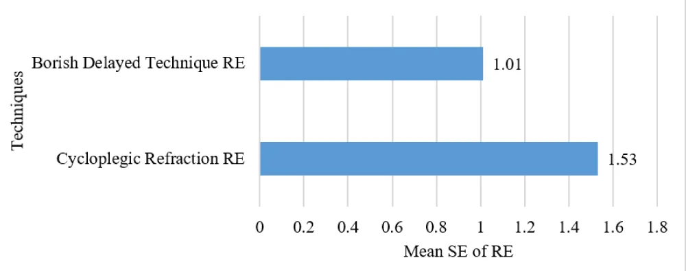
Figure 1: Comparison of of Borish Delayed Technique and Cycloplegic Refraction Mean SE of Right eye.
The rank-biserial correlation (r_rb = 0.993) and for the left eye (W = 978, p < 0.001)(Figure 2). The rank-biserial correlation (r_rb = 0.809) indicated a very large effect size, suggesting a significant difference between the two methods.
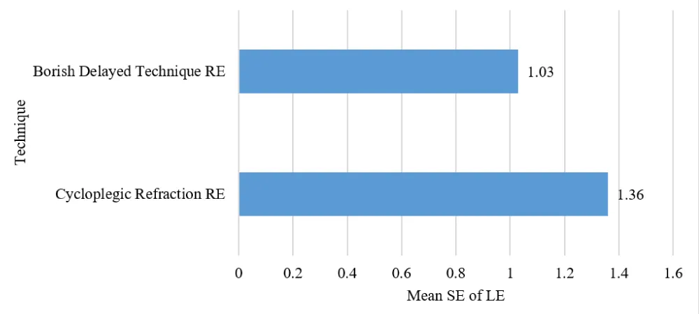
Figure 2: Comparison of of Borish Delayed Technique and Cycloplegic Refraction Mean SE of Leftt eye.
Cycloplegic refraction yielded significantly higher spherical equivalent values than the Borish delayed technique, suggesting that cycloplegia altered the refractive findings by minimizing the influence of accommodation mentioned in Table 2.
| Table 2: Comparison between Borish delayed and Cycloplegic Refraction. | |||||
| Eye | Borish Delayed Technique |
Cycloplegic Refraction |
p value | Effect Size | Interpretation |
| RE | 1.01 | 1.53 | <0.001 | 0.993 | Significant |
| LE | 1.03 | 1.36 | <0.001 | 0.809 | Significant |
The Mann–Whitney U test was performed to determine whether the difference between the Borish Delayed Technique and cycloplegic refraction varied according to gender. No statistically significant differences were observed for either the right eye (U = 294, p =0.533) or the left eye (U = 317, p =0.877). These findings indicate that gender did not significantly influence the difference between the two refractive techniques (Table 3).
| Table 3: Comparison between differences between borish delayed technique cyclo refraction with gender distribution. | ||||
| Variable | Male Median (IQR) |
Female Median (IQR) |
Mann–Whitney U | p - value |
| Difference between Borish delayed & Cyclo Refraction (RE) | 0.5 | 0.5 | 294 | 0.53 |
| Difference between Borish delayed & Cyclo Refraction (LE) | 0.5 | 0.5 | 317 | 0.88 |
Spearman’s rank correlation analysis showed no statistically significant association between age and the difference between the Borish Delayed Technique and cycloplegic refraction for the right eye (ρ = −0.104, p = 0.466) or the left eye (ρ = −0.112, p =0.435). In addition, no significant correlation was observed between the differences recorded in the right and left eyes (ρ = 0.221, p =0.121). Age was not associated with the magnitude of the difference between the two techniques (Table 4).
| Table 4: Difference between Borish delayed and cycloplegic refraction (RE) vs Difference between Borish delayed and cycloplegic refraction (LE) | |||
| Variable | Spearman's rho (r) | p value | Interpretation |
| Age Vs Difference between Borish delayed & Cyclo Refraction (RE) | -0.104 | 0.466 | Non significant-Weak negative correlation |
| Age Vs Difference between Borish delayed & Cyclo Refraction (LE) | -0.112 | 0.435 | Non significant-Weak negative correlation |
| Difference between Borish delayed & Cyclo Refraction (RE) Vs Difference between Borish delayed & Cyclo Refraction (RE) | 0.22 | 0.121 | Non significant-Weak positive correlation |
The present study compared the refractive measurements obtained using the Borish delayed technique and cycloplegic refraction in 51 children. Cycloplegic refraction yielded significantly higher spherical equivalent values than the Borish delayed technique in both eyes, indicating that it is more effective in detecting latent hyperopia. The Wilcoxon signed-rank test demonstrated a statistically significant difference between the two methods (p < 0.001), suggesting that accommodation may not be completely relaxed with the Borish delayed technique. However, statistical significance alone does not establish clinical interchangeability. The refractive values obtained by the Borish delayed technique showed good clinical agreement with cycloplegic refraction, indicating that it may serve as a useful alternative when cycloplegia is not feasible. No significant differences were observed between males and females, and age showed no significant correlation with the refractive differences. These findings support previous studies reporting that cycloplegic refraction remains the most accurate method for pediatric refractive assessment, while the Borish delayed technique can be considered a practical adjunct in selected clinical situations.
The present study demonstrated that cycloplegic refraction remains the gold standard for determining refractive errors in children, as it detected significantly higher spherical equivalent values than the Borish delayed technique. Although the Borish delayed technique provided comparable refractive measurements, it tended to underestimate latent hyperopia. Therefore, The Borish delayed technique may be considered an adjunctive approach when cycloplegic refraction is not feasible, but it should not be regarded as interchangeable with cycloplegic refraction on the basis of statistical difference testing alone. Further studies with larger sample sizes are recommended to validate these findings.
- World Health Organization. World report on vision. World Health Organization. 2019.
- GBD 2019 Blindness and Vision Impairment Collaborators, & Vision Loss Expert Group of the Global Burden of Disease Study. Causes of blindness and vision impairment in 2020 and trends over 30 years, and prevalence of avoidable blindness in relation to VISION 2020: the Right to Sight: an analysis for the Global Burden of Disease Study. The Lancet. Global health. 2021; 9(2): e144–e160. Available from: https://doi.org/10.1016/S2214-109X(20)30489-7
- Hutchinson AK, Morse CL, Hercinovic A, Cruz OA, Sprunger DT, Repka MX, et al. Pediatric Eye Evaluations Preferred Practice Pattern. Ophthalmology. 2023; 130(3): P222–P270. Available from: https://doi.org/10.1016/j.ophtha.2022.10.030
- Scheiman M, Wick B. Clinical management of binocular vision: Heterophoric, accommodative, and oculomotor anomalies (4th ed.). Wolters Kluwer. 2020.
- Gwiazda J, Thorn F, Bauer J, Held R. Emmetropization and the progression of manifest refraction in children followed from infancy to puberty. Clinical Vision Sciences. 1993; 8: 337–344.
- Ingram RM, Walker C, Wilson JM, Arnold PE, Dally S. Prediction of amblyopia and squint by means of refraction at one year. British Journal of Ophthalmology. 1986: 70(1): 12–15. Available from: https://doi.org/10.1136/bjo.70.1.12
- Benjamin WJ. (Ed.). Borish's clinical refraction (2nd ed.). ButterworthHeinemann. 2006.
- Sprunger DT, Lambert SR, Hercinovic A, Morse CL, Repka MX, Hutchinson K, Cruz, et al. Esotropia and Exotropia Preferred Practice Pattern®. Ophthalmology. 2023; 130(3): P179–P221. Available from: https://doi.org/10.1016/j.ophtha.2022.11.002
- International Agency for the Prevention of Blindness. Standard guidelines for pediatric refractive assessment. IAPB. 2021.
- Borish IM. Clinical refraction (3rd ed.). Professional Press. 1975.
- Jorge J, Queirós A, Almeida JB, Parafita MA. Retinoscopy, autorefraction and subjective refraction: Which is the best? Ophthalmic and Physiological Optics. 2005; 25(4): 370–381.
- Ministry of Health and Family Welfare. National programme for control of blindness and visual impairment: Operational guidelines. New Delhi: Government of India. 2022.
- Saxena R, Vashist P, Tandon R, Pandey RM, Bhardawaj A, Menon V, et al. Prevalence of myopia and its risk factors in urban school children in Delhi: the North India Myopia Study (NIM Study). PloS one. 2015; 10(2): e0117349. Available from: https://doi.org/10.1371/journal.pone.0117349