Figure 3
Unilateral retinitis pigmentosa: Case report and review of the literature
Denise Goodwin*, Amanda M Olsen and Karl Citek
Published: 22 August, 2019 | Volume 3 - Issue 2 | Pages: 013-19
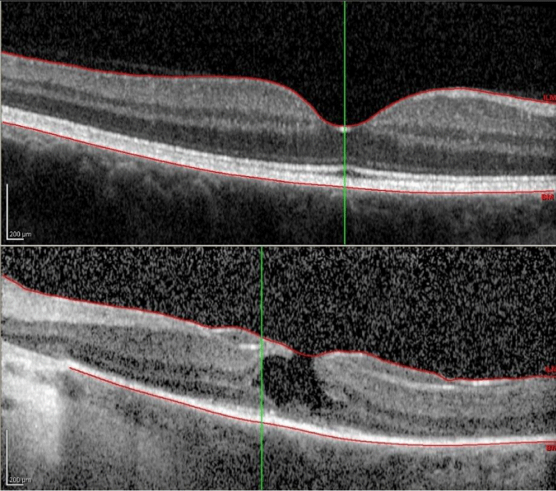
Figure 3:
Macular optical coherence tomography of the right eye (top) shows normal foveal architecture and complete photoreceptor inner segment/outer segment junction. Left eye (bottom) demonstrates a partial-thickness lamellar hole, as well as significant disruption of photoreceptor inner segment/outer segment junction, external limiting membrane, and retinal pigmented epithelium.
Read Full Article HTML DOI: 10.29328/journal.ijceo.1001021 Cite this Article Read Full Article PDF
More Images
Similar Articles
-
Detection of Ganglion Cell Loss in Preperimetric Glaucoma by Fourier-Domain Optical Coherence TomographySuneeta Dubey*, Baswati Prasanth,Lokesh Chauhan, Saptarshi Mukherjee. Detection of Ganglion Cell Loss in Preperimetric Glaucoma by Fourier-Domain Optical Coherence Tomography. . 2017 doi: 10.29328/journal.hceo.1001006; 1: 042-048
-
Intravitreal ranibizumab in the management of acute central serous ChorioretinopathyIbrahim Nawaiseh,Ahmad Halawa*,Dina Alardah. Intravitreal ranibizumab in the management of acute central serous Chorioretinopathy . . 2017 doi: 10.29328/journal.hceo.1001007; 1: 049-054
-
The management of Irvine-Gass Syndrome in a patient using Inhaler SteroidAyse Gul Kocak Altintas*,Cagri Ilhan,Mehmet Citirik. The management of Irvine-Gass Syndrome in a patient using Inhaler Steroid . . 2018 doi: 10.29328/journal.ijceo.1001011; 2: 001-005
-
Treatment of Retinal Pigment Epithelial DetachmentAyse Gul Kocak Altintas*,Cagri Ilhan. Treatment of Retinal Pigment Epithelial Detachment . . 2018 doi: 10.29328/journal.ijceo.1001013; 2: 008-014
-
Low vision due to persistent Cloquet’s canalAyse Gul Kocak Altintas*. Low vision due to persistent Cloquet’s canal . . 2018 doi: 10.29328/journal.ijceo.1001014; 2: 015-017
-
Wound architectural analysis of 1.8mm microincision cataract surgery using spectral domain OCTBenedito Antônio de Sousa, Anderson Teixeira*,Camila Salaroli,Nonato Souza,Lucy Gomes. Wound architectural analysis of 1.8mm microincision cataract surgery using spectral domain OCT. . 2019 doi: 10.29328/journal.ijceo.1001020; 3: 008-012
-
Unilateral retinitis pigmentosa: Case report and review of the literatureDenise Goodwin*,Amanda M Olsen,Karl Citek. Unilateral retinitis pigmentosa: Case report and review of the literature. . 2019 doi: 10.29328/journal.ijceo.1001021; 3: 013-19
-
Evaluation of the efficacy of transcorneal electric stimulation therapy in retinitis pigmentosa patients with electrophysiological and structural testsMualla Sahin Hamurcu*,Sema Akkan Aydogmuş,M. Sinan SARICAOĞLU. Evaluation of the efficacy of transcorneal electric stimulation therapy in retinitis pigmentosa patients with electrophysiological and structural tests. . 2020 doi: 10.29328/journal.ijceo.1001030; 4: 031-037
-
Can we predict Alzheimer’s Disease through the eye lens?Sandeep CS*,Vijayakumar N, Sukesh Kumar A. Can we predict Alzheimer’s Disease through the eye lens?. . 2020 doi: 10.29328/journal.ijceo.1001031; 4: 038-040
-
Clinical evaluation of the eye-to-visual-pathway integrity of glaucomatous neurodegeneration using 1.5T MR imaging: The integrity of glaucomatous neurodegenerationKaya N Engin*,Ulviye Yiğit#,Sibel Töreyen Bayramoğlu#,Nurten Turan Güner#,Onur Özyurt#,Kutlay Tufan#,Ahmet Ağaçhan#,Penbe Çağatay#. Clinical evaluation of the eye-to-visual-pathway integrity of glaucomatous neurodegeneration using 1.5T MR imaging: The integrity of glaucomatous neurodegeneration. . 2022 doi: 10.29328/journal.ijceo.1001042; 6: 001-006
Recently Viewed
-
Medicolegal Aspects of Deaths Due to Poisoning Occurred In Cities of Punjab and the State Of Himachal PradeshPancham Preet Kaur,Bhavish Prakash*. Medicolegal Aspects of Deaths Due to Poisoning Occurred In Cities of Punjab and the State Of Himachal Pradesh. J Forensic Sci Res. 2025: doi: 10.29328/journal.jfsr.1001087; 9: 100-103
-
Fact-finding Investigation for the Activation of the Legal-forensic Nursing Consultancy Desk at the Order of Nursing Professions of the Province of AvellinoA Masucci*,AA Di Gisi,A De Sapio. Fact-finding Investigation for the Activation of the Legal-forensic Nursing Consultancy Desk at the Order of Nursing Professions of the Province of Avellino. J Forensic Sci Res. 2025: doi: 10.29328/journal.jfsr.1001086; 9: 097-099
-
Forensic Psychology and Criminal ProfilingEze SM*,Alabi KJ,Yusuf AO,Hamzat FO,A Abdulrauf,Atoyebi AT,Lawal IA,OA Ibrahim,AY Imam-Fulani,Dare BJ. Forensic Psychology and Criminal Profiling. J Forensic Sci Res. 2025: doi: 10.29328/journal.jfsr.1001085; 9: 092-096
-
Developing an Explainable AI System for Digital Forensics: Enhancing Trust and Transparency in Flagging Events for Legal EvidenceMaruf Billah*. Developing an Explainable AI System for Digital Forensics: Enhancing Trust and Transparency in Flagging Events for Legal Evidence. J Forensic Sci Res. 2025: doi: 10.29328/journal.jfsr.1001089; 9: 109-116
-
Prediction of Stature using Percutaneous long bones of the Upper and Lower limbs among Asante and Ewe Ethnic Groups in GhanaDaniel Kobina Okwan*,Chrissie Stansie Abaidoo,Pet-Paul Wepeba,Juliet Robertson,Samuel Kwadwo Peprah Bempah,Priscilla Obeng,Ethel Akua Achiaa Domfeh,Sarah Owusu Afriyie,Thomas Kwaku Asante. Prediction of Stature using Percutaneous long bones of the Upper and Lower limbs among Asante and Ewe Ethnic Groups in Ghana. J Forensic Sci Res. 2025: doi: 10.29328/journal.jfsr.1001093; 9: 134-043
Most Viewed
-
Feasibility study of magnetic sensing for detecting single-neuron action potentialsDenis Tonini,Kai Wu,Renata Saha,Jian-Ping Wang*. Feasibility study of magnetic sensing for detecting single-neuron action potentials. Ann Biomed Sci Eng. 2022 doi: 10.29328/journal.abse.1001018; 6: 019-029
-
Evaluation of In vitro and Ex vivo Models for Studying the Effectiveness of Vaginal Drug Systems in Controlling Microbe Infections: A Systematic ReviewMohammad Hossein Karami*, Majid Abdouss*, Mandana Karami. Evaluation of In vitro and Ex vivo Models for Studying the Effectiveness of Vaginal Drug Systems in Controlling Microbe Infections: A Systematic Review. Clin J Obstet Gynecol. 2023 doi: 10.29328/journal.cjog.1001151; 6: 201-215
-
Prospective Coronavirus Liver Effects: Available KnowledgeAvishek Mandal*. Prospective Coronavirus Liver Effects: Available Knowledge. Ann Clin Gastroenterol Hepatol. 2023 doi: 10.29328/journal.acgh.1001039; 7: 001-010
-
Causal Link between Human Blood Metabolites and Asthma: An Investigation Using Mendelian RandomizationYong-Qing Zhu, Xiao-Yan Meng, Jing-Hua Yang*. Causal Link between Human Blood Metabolites and Asthma: An Investigation Using Mendelian Randomization. Arch Asthma Allergy Immunol. 2023 doi: 10.29328/journal.aaai.1001032; 7: 012-022
-
An algorithm to safely manage oral food challenge in an office-based setting for children with multiple food allergiesNathalie Cottel,Aïcha Dieme,Véronique Orcel,Yannick Chantran,Mélisande Bourgoin-Heck,Jocelyne Just. An algorithm to safely manage oral food challenge in an office-based setting for children with multiple food allergies. Arch Asthma Allergy Immunol. 2021 doi: 10.29328/journal.aaai.1001027; 5: 030-037

HSPI: We're glad you're here. Please click "create a new Query" if you are a new visitor to our website and need further information from us.
If you are already a member of our network and need to keep track of any developments regarding a question you have already submitted, click "take me to my Query."


