More Info
Author's satisfaction with
- Friendly and hassle-free publication process
- Less production time of articles
- Constructive peer-review
- Enhancing journal reputation
- Regular feedback system
- Quick response to authors' queries
Recently Viewed
Most Viewed
Clinical Images
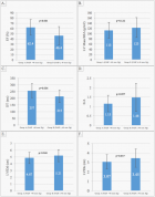
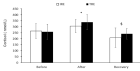
Table of Contents

Prospective Clinical Study to Find out Epidemiology of Xerophthalmia in Children in a Tertiary Care Centre in India
Published on: 29th December, 2017
OCLC Number/Unique Identifier: 7355977840
Objective: To study the epidemiology of xerophthalmia in children 2-6 years of age in North India.
Methods: A prospective clinical study was done at two tertiary care centers of North India between 2010 to 2016, Cases were selected from routine OPD and children less than 6 years of age were examined by an ophthalmologist. Diagnosis and classification of Xerophthalmia was done according to WHO classification. All the data recording demographic profile, socioeconomic status, other health problems etc were recorded in a fixed proforma. Data was analyzed by SPSS version 16.
Findings: Two thousand nine hundred forty six cases were included in the study after satisfying inclusion and exclusion criteria. The prevalence of night blindness was estimated to be 2.93% (95% Confidence Interval [CI]: 2.53-3.33) among children between 2 and 6 years of age. Xerophthalmia prevalence was 4.43% (95% CI: 4.19-4.67). Prevalence was more in girls than boys and higher in low socioeconomic status.
Conclusion: Vitamin A deficiency is recognized to be a severe public health problem leading to corneal opacity and childhood blindness in most of the areas of North India
Neuro-ophthalmological emergency disorders: A general view
Published on: 27th December, 2017
OCLC Number/Unique Identifier: 7355943544
Neuro-ophthalmological emergency disorders usually occur with symptoms of visual loss, diplopia, ocular motility impairment and anisocoria. In this mini-review, we aim to take look the common neuro-ophthalmological emergency disorders. The delayed diagnosis of the neuro-ophthalmological emergencies puts the patient at risk of death or blindness. If these are well-known, the discrimination and management of these emergency conditions will be easier.Introduction
The Role of Omega-3 Essential Fatty Acids in Dry Eye Disease
Published on: 22nd December, 2017
OCLC Number/Unique Identifier: 7355944105
One of every four visits to eye care professionals is for dry eye disease which affects an estimated 7-34% of Americans [1]. Knowledge regarding etiology and treatments has advanced exponentially in the last 20 years.
Intravitreal ranibizumab in the management of acute central serous Chorioretinopathy
Published on: 24th November, 2017
OCLC Number/Unique Identifier: 7317652738
Purpose: To evaluate the efficacy of ranibizumab in hastening the recovery of acute CSCR when given immediately at time of diagnosis.
Methods: In This retrospective case series, a total of 72 patients diagnosed with acute CSCR where reviewed, of which 63 received Ranibizumab at presentation. The patients were evaluated using Best corrected visual acuity, Ophthalmic examination, Optical coherence tomography (OCT) and fluorescein angiography, in addition to indocyanine green angiography and OCT angiography in some cases, at presentation, one week, one month and two months’ post injection.
Results: From the total 72 patients diagnosed with acute CSCR, 63 of them received intravitreal ranibizumab and the remaining 9 patients preferred to go for observation. The mean age of patients was 41.2 year old. The ratio of male to female was 8:1. The mean BCVA at presentation was 6/15 on Snellen chart. All patients who received ranibizumab injection showed an improvement after 1 week, with a mean improvement in BCVA of two lines. Of them, 43 patients were back to BCVA of 6/6 after 2 months and showed complete resolution of sub retinal fluid. The remaining 20 patients showed an additional mean of improvement of one line (over the previous two lines) after the 2 months.
Conclusion: Intravitreal ranibizumab hasten the recovery of both the BCVA and central macular thickness on OCT in acute CSCR when given immediately at presentation.
Detection of Ganglion Cell Loss in Preperimetric Glaucoma by Fourier-Domain Optical Coherence Tomography
Published on: 24th October, 2017
OCLC Number/Unique Identifier: 7317651893
Background: Glaucoma is a multi-factorial optic neuropathy characterized by a loss of retinal ganglion cells with subsequent loss of the retinal nerve fibers ultimately resulting in visual impairment. The macula region has a high density of retinal ganglion cells thereby being a likely region to detect early cell loss .Since glaucoma affects mainly the inner layers of the retina, Ganglion Cell Complex (GCC) mapping can help to detect glaucomatous damage early as compared to the total retinal thickness.
Purpose: To map GCC thickness and average Macular Retinal (MR) thickness with high-speed Fourier-Domain Optical Coherence Tomography (FD-OCT) and correlate it with the Retinal Nerve fiber layer (RNFL) thickness in preperimetric glaucoma.
Design: Observational cross-sectional study.
Methods: Forty four eyes diagnosed as preperimetric glaucoma were studied. GCC, MR thickness and RNFL thickness was mapped using the RTVue FD-OCT system. The GCC thickness map, the deviation map and the significance map were obtained in all cases. Average GCC thickness and MR thickness were correlated with the RNFL thickness.
Results: Average GCC of patients was 85.99±6.9 µm. There was GCC loss in 35 (87.5%) eyes which correlated well with areas of RNFL loss (r=0.408, p<0.001). Nine (22.5%) eyes were seen to have decreased MR thickness. GCC loss correlated well with the loss of average RNFL thickness and MR thickness. Further GCC loss was also seen in 23 (74.19 %) eyes with a normal MR thickness.
Conclusion: GCC analysis may prove to be a robust diagnostic parameter and is complementary to RNFL analysis in preperimetric glaucoma.
Intravitreal Ranibizumab/ Lucentis (IVTL) injections in Glaucoma patients-Intraocular Pressure (IOP) elevation and the use of Anterior Chamber Paracentesis (ACP)
Published on: 20th September, 2017
OCLC Number/Unique Identifier: 7317592360
Purpose
• To assess the short term effects of intravitreal Lucentis (IVTL) on intraocular pressure in patients with ocular hypertension and glaucoma
• To determine rate of anterior chamber paracentesis (ACP) required post-injection according to departmental protocol
Methods
This was a prospective, observational study carried out between August 2011 and February 2012 in the Department of Ophthalmology, Maidstone Hospital. 24 participants (13 female, 11 male) with established ocular hypertension (OHT) or glaucoma were chosen from a cohort of patients receiving intravitreal (IVTL) Ranibizumab (Lucentis) treatment for wet age related macular degeneration (wARMD). Apraclonidine 1% was given pre-injection, and baseline IOP was measured 30 min. after this, just before IVTL. IOP was measured at baseline, within 1 min of injection, 5 min, 15 min, 30 min up to 60min following a single IVTL treatment.
Anterior paracentesis was performed if:
• Immediate post injection IOP > 50mm Hg and OHT
• Immediate post injection IOP > 40 mm Hg and there was evidence of disc damage only
• Immediate post injection IOP > 30mm Hg with evidence of disc damage and visual field loss
Results
79.2% had diagnosed disc damage and visual field loss (glaucoma); 12.5% had disc damage only (pre-perimetric glaucoma), whereas the remaining 8.3% had no evidence of disc damage or visual field loss i.e. ocular hypertension (OHT).
Administration of Apraclonidine 1% prior to IVTL did not cause a statistically significant IOP reduction in patients with OHT and glaucoma (paired Student’s t-test P = 0.368). Immediately post injection, mean IOP was 41.54mm Hg (SD 14.1, 95% CI 37.20 to 45.88; Paired T test results P <0.0001,) which confirmed a statistically significant difference between baseline and immediate post injection IOP.
13 out of 24 (58%) of the study patients required anterior chamber paracentesis (ACP) post IVTL according to our devised protocol. There was no statistically significant difference in baseline IOP between the paracentesis and non-paracentesis groups (p=0.4). The presence of a bleb post injection had no statistically significant bearing on immediate post intravitreal IOP (p=0.3).
ACP performed at 1min restored IOP to a safer level at 5min in all cases thus treated.
Conclusions
IVTL appears to cause a significant but transient rise in IOP which is reduced after a mean time of 5 minutes. Although the clinical significance of this IOP spike is still unknown, extreme care must be taken in patients with ocular hypertension and glaucoma particularly those with established disc damage and visual field loss. Apraclonidine 1% appears to have a limited role in the prophylactic lowering of IOP pre-injection. The authors propose the use of the formulated anterior chamber paracentesis protocol for IOP management in patients with OHT and glaucoma receiving intravitreal anti-VEGF treatment.
Mitomycin-C Use and Complications in Ophthalmology
Published on: 28th June, 2017
OCLC Number/Unique Identifier: 7317596987
Mitomycin-C, first found its way into ophthalmic use in 1969, in Japan, where recurrent pterygia were successfully treated with the drug which is an antineoplastic / antibiotic agent isolated from the soil bacterium Streptomyces caespitosus [1]. It is an anti-metabolite with anti-proliferative effect on cells showing the highest rate of mitosis by inhibiting DNA synthesis and interferes with RNA transcription and protein synthesis [2].CLINICAL USES
Efficacy of early Hyperbaric Oxygen Therapy in Central Retinal Artery Occlusion
Published on: 20th June, 2017
OCLC Number/Unique Identifier: 7317650795
A 60-year-old woman had central retinal artery occlusion (CRAO) presented to the ophthalmology department with a sudden, painless loss of vision. Her initial visual acuity was light perception and she treated with hyperbaric oxygen therapy (HBOT) 4 hours after the development of visual symptoms. Systemic risk factors were not found so she was diagnosed as having idiopathic CRAO. Her vision improved from light perception to 20/50 after the HBOT. Any complications such as neovascularization were not developed until the last follow up visit of 8th months.
Theory and Experiments. (+) Add Reading Glasses to Prevent Myopia
Published on: 20th February, 2017
OCLC Number/Unique Identifier: 7317576307
Basic control theory equations are developed showing conventional exponential system response of refraction vs. time R (t) with a characteristic system time constant, in response to a step change of near work environmental conditions. Details from preliminary experimental design using reading glasses at the U.S. Naval Academy at Annapolis are discussed. The conclusion is that (+) add lenses, used as reading glasses during study, can prevent the development of myopia for college students in pilot training.
A Comparative Study of Anatomic and Functional Outcomes of Two Surgical Techniques of Cataract at Lome
Published on: 17th February, 2017
OCLC Number/Unique Identifier: 7317592098
Aim: To compare the anatomical and functional outcomes of cataract surgery with manual small incision cataract surgery (MSICS) to those of extracapsular cataract extraction (ECCE) in Lome.
Patients and Methods: A prospective study involved two groups of patients who underwent ECCE (group 1) and MSICS (group 2) by the same surgeon in the same conditions in different periods. Complications and visual results to the 45th postoperative day were compared.
Results: At the 45th postoperative day, 60% of operated eyes of the ECCE group (G1) and 83.9% in the group of MSICS (G2) had uncorrected visual acuity greater than or equal to 3/10. Through the pinhole, these proportions increased to 73.3% for G1 and 92.2% for G2. Visual acuity was less than 1/10 in 4.4% for G1 and 1.1% for G2. The vitreous loss was observed in proportions of 3.8% for G1 and 3.3% for G2. During follow-up, the three main early postoperative complications were inflammation (13.9%), corneal edema (13.3%), and the pigment dispersion (7.2%) in G1 and corneal edema (9.4%), pigment dispersion (8.3%) and hypertonia (6.6%) in G 2.
Conclusion: Two cataract extraction techniques offer the same level of safety in intraoperative period. However, MSICS has certain advantages over the ECCE and would be an alternative technique in developing countries.
Contact
Select by Volume & Issue
Most Viewed Keywords
- Operative surgical procedures
- Endoscopic
- Cardiovascular risk
- Obesity
- Assisted reproduction
- Dynamic panel data
- Armored scale insect
- ZYMV
- Deep venous collateral angiography
- β-thalassemia major
- CYP27A1 mutation
- Telemedicine
- Municipal solid waste
- Relative bone density
- BMI
- Groundwater
- Web browsers
- Total bronchoalveolar lavage
- Bariatric surgery
- Priapism
University/Institution
Select and search by University/Institution.
Articles by Country
Select and search by country to get related articles.


Testmonials

I would like to thank JPRA for taking this decision. I understand the effort it represents for you. I'm truly happy to have the paper published in JPRA. And I'll certainly consider JPRA for my next publications as I was satisfied of the service provided, the efficiency and promptness of the interactions we had.
Emmanuel BUSATO
Publishing with the International Journal of Clinical and Experimental Ophthalmology was a rewarding experience as review process was thorough and brisk. Their visibility online is second to none as their published articles appear in all search engines. I will encourage researchers to publish with them.
Elizabeth Awoyesuku
“The choice to submit the forensic case study to the Journal of Addiction Therapy and Research was dictated by the match between the content and the potential readership. The publication process proved to be expedient and we were provided with constructive feedback from reviewers. The final article layout is attractive and conforms to standards. All-in-all, it has been a rewarding process.”
Elisabeth H Wiig
Archives of Vascular Medicine is one of the top class journal for vascular medicine with highly interesting topics. You did a professional and great Job!
Elias Noory
Thank you very much. I think the review process and all of what concerns the administration of the publication concerning our paper has been excellent. The nice and quick answers have been very good I think.
Doris Nilsson
Journal of Pulmonary and Respiratory Research is good journal for respiratory research purposes. It takes 2-3 weeks maximum for review of the manuscript to get published and any corrections to be made in the manuscript. It needs good articles and studies to get publish in the respiratory medicine. I am really glad that this journal editors helped me to get my case report published.
Divya Khanduja
Thanks you and your colleague for the great help for our publication. You always provide prompt responses and high quality of service. I am so happy to have you working with me. Thanks again!
Diana (Ding) Dai
Service and process were excellent as was the “look” of the article when published.
Deane Waldman
Great, thank you! It was very efficient working w/ your group. Very thorough reviews (i.e., plagiarism, peer, etc.). Would certainly recommend that future authors consider working w/ your group.
David W Brett
Your services are very good
Chukwuka Ireju Onyinye
I very much appreciate the humanitarian services provided in my stead by this journal/publisher. It exhibits total absence of editorial impertinence. As an Author, I have been guided to have a fruitful experience. The editorial care is highly commendable.
Chrysanthus Chukwuma
"An amazing experience with the Journal of Advanced Pediatrics and Child Health. Very fast blind review with pertinent corrections and suggestions. I highly recommand both the journal and the editor."
Chaimae Khairoun
The submission is very easy and the time from submission to response from the reviewers is short. Correspondence with the journal is nice and rapid.
Catrin Henriksson
The Clinical Journal of Obstetrics and Gynecology is an open access journal focused on scientific knowledge publication with emphasis laid on the fields of Gynecology and Obstetrics. Their services toward us have been encouraging through their kindness and respect. Great consideration has been given to us as young budding researchers and we are very grateful for this.
Carole Assontsa
During the process your positive communication, prompt feedback and professional approach is very highly appreciated. We would like to thank you very much for your support.
Can Vuran
I do appreciate for your service including submission, analysis, review, editorial and publishing process. I believe these esteemed journal enlighten the science with its high-quality personel.
Bora Uysal
I am very much pleased with the fast track publication by your reputed journal's editorial team. It is really helpful for researchers like me from developing nations. I strongly recommend your journal for publication.
Badri Kumar Gupta
It has been a fabulous journey writing articles for your journal because of the encouragement you people provide for writers from developing nations like India. Kindly continue the same. Looking forward for a long term association.
Badareesh Lakshminarayana
Many thanks for publishing my article in your great journal and the friendly and hassle-free publication process, the constructive peer-review, the regular feedback system, and the Quick response to any queries.
Azab Elsayed Azab
I would like to thank this journal for publishing my Research Article. Something I really appreciate about this journal is, they did not take much time from the day of Submission to the publishing date. Looking forward to have more publications in future.
Ayush Chandra
Submission of paper was smooth, the review process was fast. I had excellent communication and on time response from the editor.
Ayokunle Dada
Your service is very good and fast reply, also your service understand our situation and support us to publication our articles.
Ayman M Abu Mustafa
Really good service with prompt response. Looking forward to having long lasting relationship with your journal
Avishek Bagchi
Your service is excellent. Processing and editing were very fast. I hope to publish more of my works in your journal.
Ausraful Islam
I wanna to thank Clinical Journal of Nursing Care and Practice for its effort to review and publish my manuscript. This is reputable journal. Thank you!
Atsedemariam Andualem
“It was a delightful experience publishing my manuscript with the Clinical Journal of Obstetrics and Gynecology. They offered me lots of opportunities I never had from most publishing houses and their prompt services are greatly appreciated.”
Asafo Jones
I hope to ability to make some new investigation and publish in Your Company in future.
Artur Stopyra
I like the quality of the print & overall service. The paper looks quite impressive. Hope this will attract interested readers. All of you have our best wishes for continued success.
Arshad Khan
Your big support from researchers around the world is the best appreciation from your scientific teams. We believe that there should be no barrier in science and you make it real and this motto come true.
Arefhosseinir Rafi
Your journal co-operation is very appreciable and motivational. I am really thankful to your journal and team members for the motivation and collaboration to publish my work.
Assistant Professor, UCLAS Uttaranchal University, Dehradun, India
Archna Dhasmana
I am glad to submit the article to Heighten Science Publications as it has a very smooth and fast peer-review process, which enables the researchers to communicate their work on time.
Anupam M
This is to specify that I have had an extensive and detailed interaction with the Editorial team of Annals of Clinical Gastroenterology and Hepatology, USA, lasting over a significant period of time. My interaction has been extremely pleasant, especially with Ms Allie Smith, as I find the communication quite inspiring and crystal clear. The attitude of aforesaid individuals is quite helpful and guiding in pertinent instances. It has been a commemorative journey so far working with the Journal and I hope that the symbiosis will continue, evolve and flourish in the forthcoming years. I wish the journal, related personnel and aforementioned individuals a fruitful, successful run.
New Delhi, India
Anubha Bajaj
We appreciate the fact that you decided to give us full waiver for the applicable charges and approve the final version. You did an excellent job preparing the PDF version. Of course we will consider your magazine for our future submissions and we will pay the applicable fees then.
Anna Dionysopoulou
''Co-operation of Archives of Surgery and Clinical Research journal is appreciable. I'm impressed at the promptness of the publishing staff and the professionalism displayed. Thank you very much for your support, help and encouragement.''
Anıl Gokce
Congratulations for the excellence of your journal and high quality of its publications.
Angel MARTIN CASTELLANOS
The service from the journal staff has been excellent.
Andy Smith
I was very pleased with the quick editorial process. We are sure that our paper will have great visibility, among other things due to its open access. We believe in science accessible to all.
Anderson Fernando de Souza
It was a great experience publishing through JCICM. The article has reached out to several institutions. Appreciate your professional work. Hope to work with you again
Anas Wardeh
Publishing an article is a long process, but working with your publication department made things go smoothly, even though the process took exactly 5 months from the time of submitting the article till the time I have favourable response, the missing part is the peer review details, which is essential in self auditing and future improvement, overall experience was excellent giving your understanding of the situation of lack of financial institution support.
Anas Diab
I think that Heighpubs very good. You are very helpful. Thank you for everything.
Ana Ribeiro
Regarding to be services, we note that are work with high standards of professionalism translated into quick response, efficiency which makes communication accessible. Furthermore, I believe to be much inviting for the submission of future works for publication purposes.
Amélia João Alice Nkutxi
I would like to mention that I had a wonderful experience working with HSPI. The whole process right from manuscript submission to peer review till the publication of the article was very prompt & efficient. I wish you good luck for the future.
Amarjeet Gambhir
Once I submitted the manuscript, the response time of the reviewers was very fast. The fine-tuning of the galley proof was likewise prompt. I believe the journal provide a valuable outlet to disseminate physical rehabilitation scientific knowledge to the clinical community. Respectfully. Dr. Alon
Alon
We really appreciate and thanks the full waiver you provide for our article. We happy to publish our paper in your journal. Thank you very much for your good support and services.
Ali Abusafia
It was a real pleasure working with your team. The review was done fast, and it was very clear, the editing was flawless, the article was published quickly compared to other journals, and everyone was understanding and helpful. I will gladly recommend this journal to my acquaintances in academia.
Alexandra Cozma
To the editorial team at HSPI and the Journal of Clinical Nephrology: Thank you so much for your hard work and collaboration in bringing our article to life. Your staff was responsive, flexible, and communicative and made the process smooth and easy. Thank you!
Alejandro Munoz
Dear colleagues! I am satisfied with our cooperation with you. Your service is at a high level. I hope for a future relationship. Let me know if I can get a paper version of the magazine with my articles from you. I see them on the Internet.
Aksenov V.V
"This is my first time publishing with the journal/publisher. I am impressed at the promptness of the publishing staff and the professionalism displayed. Thank you for encouraging young researchers like me!"
Ajite Kayode
I want to thank you for our collaboration. You were fast and effective with a positive spirit of teamwork. I am truly excited from our collaboration. You were like always fast, efficient and accurate. I hope that in the near future we will collaborate again.
Aikaterini Solomou
In my opinion, you provide a very fast and practical service.
Ahmet Eroglu
Great, We are too comfortable with the process including the peer review process and quality. But, the journal should be indexed in different databases such scopus.
Afework Edmealem
We really appreciate your efforts towards our article, the professional way you handle our request for exemption from charges. It was a great honor for us to publish in your magazine.
Achraf elbakkaly
I really liked the ease of submitting my manuscript in the HSPI journal. Further, the peer review was timely completed and I was communicated the final decision on my manuscript within 10 days of submission which is really appreciable. I strongly recommend all the scientists and researchers to submit their work in this journal”
Abu Bashar
My candid opinion is that the service you render is second to none. My favourite part is the prompt response to issue, really i value that.
Abiodun Akanbi Adeogun
Thank you very much for accepting our manuscript in your journal “International Journal of Clinical Virology”. We are very thankful to the esteemed team for timely response and quick review process. The editorial team of International Journal of Clinical Virology is too cooperative and well-mannered during the publication process. We are hopeful to publish many quality papers in your journal and I suggest the International Journal of Clinical Virology to all of my colleagues, researchers and friends to publish their research here.
Abdul Baset
I, Muhammad Sarwar Khan, am serving as Editor on Archives of Biotechnology and Biomedicine (ABB). I submitted an editorial titled, 'Edible vaccines to combat Infectious Bursal Disease of poultry' for publication in ABB. After submitting the manuscript; the services rendered by the management and technical personnel to handle and process the manuscript were marvelous. Plagiarism report was shared with me with complements before reviewers' comments, All steps including article processing and service charges were well taken care of keeping in view the author's interest/preference. All together, it was an encouraging and wonderful experience working with ABB personnel.
University of Agriculture, Pakistan
Muhammad Sarwar Khan
Your journal has accomplished its intended mission of providing very effective and efficient goals in dealing with submissions, conducting the reviewing process and in publishing accepted manuscripts in a timely manner. Keep up the great work and services that you provide.
University of Jacqmar, Inc., USA
John St. Cyr
I am to express my view that Heighten Science Publications are reliable quick even after peer review process. I hope and wish the publications will go a long way in disseminating science to many interested in scientific research.
College of Fisheries, CAU(I), Tripura, India
Ajit Kumar Roy
The Journal Clinical Nephrology provides a good opportunity for readers to stay updated in the field of clinical nephrology. Additionally - it provides a good opportunity for authors to publish their work. 1. Publication of the accepted manuscripts is sufficiently rapid. 2. The trust factor between the journal and me, as an author, is very important and well preserved. 3. Peer review process very rapid and effective.
Assaf Harofeh Medical Center, Israel
Leonid Feldman
In 2017, I submitted a manuscript to the journal Archives of Biotechnology and Biomedicine belonging to Heighten Science Publications Corporation. Within one week I already received the response from the editor. All processing steps were really fast so in terms of a speedy publication I can particularly recommend the journal Archives of Biotechnology and Biomedicine. The responsible contact person of the journal was always available, which gives a trustworthy impression to the author. Also the peer review process was clear and constructive. So from my experience with Heighten Science Publications Corporation I can recommend publishing there.
University of Tubingen, Germany
Yvonne Mast
We thank to the heighten science family, who speed up the publication of our article and provide every support.
Mehmet Besir
The services of the journal were excellent. The most important thing for an author is the speed of the peer review which was really fast here. They returned in a few days and immediately replied all of my questions. I want to refer this platform to all scholars. Many thanks.
Eastern Mediterranean University, Cyprus
Zehra Guchan TOPCU
Thank you for your attitude and support. I am sincerely grateful to you and the entire staff of the magazine for the high professionalism and fast quality work. Thank you very much!
USA
Igor Klepikov
Thank you and your company for effective support of authors which are very much dependable on the funds gambling for science in the different countries of our huge and unpredictable world. We are doing our work and should rely on a teams like Galley Proof-HSPC. Great success to all of you for the 2019th! Be well all the year long.
Russia
Victor V Apollonov
The editorial process was quickly done. The galley proof was sent within a week after being accepted for publication. The editorial team was very helpful and responded promptly.
India
Rohit Kulshrestha
Publishing with the International Journal of Clinical and Experimental Ophthalmology was a rewarding experience as review process was thorough and brisk. Their visibility online is second to none as their published articles appear in all search engines. I will encourage researchers to publish with them.
University of Port Harcourt Teaching Hospital, Nigeria
Dr. Elizabeth A Awoyesuku
"It was a pleasure to work with the editorial team of the journal on the submission of the manuscript. The team was professional, fast, and to the point".
NC A&T State University, USA
Moran Sciamama-Saghiv
Submission of paper was smooth, the review process was fast. I had excellent communication and on time response from the editor.
Ekiti State University Teaching Hospital, Nigeria
Ayokunle Dada
I am delighted and satisfied with. Heighten Science Publications as my manuscript was thoroughly assessed and published on time without delay. Keep up the good work.
Ido-Ekiti/Afe Babalola University, Nigeria
Dr. Shuaib Kayode Aremu
"This is my first time publishing with the journal/publisher. I am impressed at the promptness of the publishing staff and the professionalism displayed. Thank you for encouraging young researchers like me!"
Ekiti State University, Nigeria
Adebukola Ajite
I wanna to thank clinical journal of nursing care and practice for its effort to review and publish my manuscript. This is reputable journal. Thank you!
Wollo University, Ethiopia
Atsedemariam Andualem
We appreciate your approach to scholars and will encourage you to collaborate with your organization, which includes interesting and different medical journals. With the best wishes of success, creativity and joy in life, prosperity in the medical field.
Ivano- Frankivsk National Medical University, Ukraine
Nataliya Kitsera
Thank you very much for your support and encouragement. I am truly impressed by your tolerance and support. Thank you very much
Diaverum: PADC, Jeddah, Saudi Arabia
Nasrulla Abutaleb
You are such a nice person. Your journal co-operation is very appreciable and motivational.
Department of Biotechnology, Uttaranchal college of Applied and Life Sciences, Uttaranchal University, Dehradun, Uttarakhand, India
Archna Dhasmana
“Mobile apps and wearable technology are becoming ubiquitous in our environment. Their integration with healthcare delivery is just beginning to take shape. The early results are promising and the possibilities great."
BS, PharmD., MBA, CPHIMS, FHIMSS, Adjunct Professor, Global Healthcare Management, MCPHS University, Chief Strategy Offi cer, MedicaSoft, Senior Advisor, National Health IT (NHIT) Collaborative for Underserved, New York HIMSS, National Liaison, Health 2.0 Boston, Past Chair, Chair Innovation, USA
Helen Figge
“The choice to submit the forensic case study to the Journal of Addiction Therapy and Research was dictated by the match between the content and the potential readership. The publication process proved to be expedient and we were provided with constructive feedback from reviewers. The final article layout is attractive and conforms to standards. All-in-all, it has been a rewarding process.”
Ph.D, Boston University Department of Communication Sciences and Disorders and Knowledge Research Institute, Inc., 2131 Reflection Bay Drive, Arlington, Texas 76013, USA
Elisabeth H. Wiig
The service is nice and the time of processing the application is fast.
Department of Neurosurgery, Queen Elizabeth Hospital, Hong Kong
Long Ching
Your service is very good and fast reply, Also your service understand our situation and support us to publication our articles.
Palestine College of Nursing, Khan Younis, Gaza Strip, Palestine
Ayman M Abu Mustafa
“It was a delightful experience publishing my manuscript with the Clinical Journal of Obstetrics and Gynecology. They offered me lots of opportunities I never had from most publishing houses and their prompt services are greatly appreciated.”
Department of Agricultural Economics, Agribusiness and Extension, Kwame Nkrumah University of Science and Technology, Kumasi, Ghana
Akowuah Jones Asafo
Related Journals

HSPI: We're glad you're here. Please click "create a new Query" if you are a new visitor to our website and need further information from us.
If you are already a member of our network and need to keep track of any developments regarding a question you have already submitted, click "take me to my Query."