More Info
Author's satisfaction with
- Friendly and hassle-free publication process
- Less production time of articles
- Constructive peer-review
- Enhancing journal reputation
- Regular feedback system
- Quick response to authors' queries
Recently Viewed
Most Viewed
Clinical Images
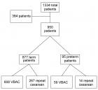
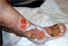
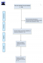
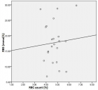
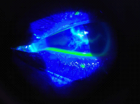
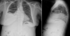
Table of Contents

The evaluation of bandage soft contact lenses as a primary treatment for traumatic corneal abrasions
Published on: 25th May, 2020
OCLC Number/Unique Identifier: 8605482786
Background: Corneal abrasions are a common result of eye trauma. Corneal injuries are very common in both the adult and pediatric population and account for a significant proportion of the workload of most emergency departments. Although abrasion heals well with preservative treatment, it still causes pain and job lost. The abrasion result from the scrabble of the corneal epithelium. These injuries cause pain, tearing, lids spasm, light scare, foreign body sensation, decreased visual acuity/blurring, and a gritty feeling. The light, friction & wink was worse the condition. Most abrasion cure within 24-27 hours and seldom proceed to erosion or infection. The study aims to use bandage soft contact lens [BSCL] as a primary treatment for traumatic corneal abrasion [TCA] instead of traditionally use pressure patch [PP].
Patients and methods: The present prospective study has been conducted on 50 patients attending the out-patient department of ophthalmology in an Alyarmouk teaching hospital for six months after taking ethical permission. Before subjecting the patient to the treatment of bandage soft contact lens therapy, a detailed clinical history and thorough local examination have been done. A history indicating the occurrence of recent ocular trauma followed by severe pain, redness, lids spasm, photophobia, and tearing of the involved eye is suggestive of a corneal abrasion. Always we ask about contact lens wear as this can complicate the presence of an abrasion. To confirm the diagnosis of traumatic corneal abrasion we examine the cornea by slit-lamp under cobalt-blue filtered light after the application of tetracaine eye drops & fluorescein strips. The treatment of 50 consecutive patients presenting with traumatic corneal abrasion has been treated with anesthetic eye drop (tetracaine 0.5%) to relieve pain and lids spasm, antibiotic eye drop (ofloxacin 0.3%), therapeutic bandage soft contact lens was applied to provide pain relief and once again act as a splint to promote epithelial healing, then visual acuity was measured by Snellen chart, a cycloplegic eye drop (cyclopentolate 1%) was applied to relieve ciliary spasm & then preservative-free lubricant eye drop were applied lastly. This criterion dramatically relieves most, if not all of the pain the patient may be experiencing (which is a big plus for the patient and earns instantaneous trust), but it also allows the patient to return to work/school or any other daily activities. Patients have been evaluated after 24hours, 72hours and after 1week regarding pain, visual acuity, and complications. Though pressure patch [PP] occasionally advice in abrasion therapy, it does not assist and may prevent recovery. Employ the protective eyewear can preclude the traumatic corneal abrasion.
Results: A total of 50 cases were enrolled in our study during the study period of 6 months. Out of 50 patients, there were 30males and 20 females and the male/female ratio was 3:2. The patient’s age was ranged from 5-35years. The commonest cause of injury was direct minor trauma (80% of cases), with cosmetic & optical contact lenses related problems accounting for 20% of presentations, visual acuity was documented correctly in 90% of adult and pediatric group and difficult to documented in children less than 6-year-old 10%. Traumatic corneal abrasion treated with bandage soft contact lens has an apparent advantage over the traditional pressure patch in terms of reduced pain, speedier healing, and an advantage of faster rehabilitation, facilitation epithelial healing, and proper surface hydration. Evaluation of pain revealed sufficient comfort with this regimen, allowing 45 patients (90%) to go back immediately to their occupations. Moreover, visual function is retained without any complication. Healing of the traumatic corneal abrasion occurred within 1 to 3 days in all patients, with minimal or no pain. The infection did not occur at the time of the follow up. We remove the bandage soft contact lens after 1 week to allow epithelial migration and attachment without the interference of the shearing forces of the upper lid.
Conclusion: The use of bandage soft contact lens as a primary treatment for a traumatic corneal abrasion is a safe and effective method with anesthetic eye drop (tetracaine 0.5%), antibiotic eye drop (ofloxacin 0. 3%), cycloplegic eye drop (cyclopentolate 1%), preservative-free lubricant drop instead of traditionally pressure patch. Bandage soft contact lens causes dramatic improvement from pain, lid spasm, tearing & visual function is retained without any complication, and patients can immediately resume their regular activities.
Can we predict Alzheimer’s Disease through the eye lens?
Published on: 22nd May, 2020
OCLC Number/Unique Identifier: 8601971854
Alzheimer’s Disease (AD) is a common dementia problem of the old population. The two main hallmarks of AD are tau protein and amyloid-beta protein. The relevant investigations on AD suggest that these proteins are also seen in the eye. There are many tests and imaging modalities are used for AD diagnosis. But these techniques are still unable to predict the disease effectively. In this regard, the lens of the eye may help in diagnosing AD. Therefore, a reliable technique for measuring the lens or retina must be selected. In this paper, we focus on the different types of retinal diseases occur in AD patients and the use of the Optical Coherence Tomography (OCT) technique is used for diagnosing AD.
Evaluation of the efficacy of transcorneal electric stimulation therapy in retinitis pigmentosa patients with electrophysiological and structural tests
Published on: 20th May, 2020
OCLC Number/Unique Identifier: 8604562702
A Statement of significance: This study shows that the effect of transcorneal electrical stimulation (TES) therapy as a stimulator device in retinitis pigmentosa (RP)patients with have a significant increase in visual acuity and shortening of p100 latency in pattern visual evoked potential (pVEP) test during 3 months follow up.
Purpose: To assess the safety and efficacy of TES therapy with electrophysiological and structural tests in RP patients.
Methods: Thirty four eyes of 17 RP patients were included in the study. Initial examination included best corrected visual acuity (BCVA) and visual field (VF) test (Humphrey). Central macular thickness (CMT), retinal nerve fiber layer thickness (RNFLT) and choroidal thickness (CT) were measured with using swept-source optical coherence tomography (OCT). The patients were tested by Metrovision brand monpack model visual eletrophysiology device for pVEP and flash electroretinogram (fERG) tests. Patients were seen 12 times during 3 months: initial visit for screening and weekly visits for TES. All tests were repeated 3 times. The results of pre and post TES therapy were compared.
Results: Patients’ baseline BCVA was 0,34 ± 0,22. The increase in the last visit BCVA was significant (p : .001) and it was 0.50 ± 0.29. The difference between CMT, RNLF and CT pre and post TES therapy were not significant (p > .05). The mean latencies of the 120’ pattern p100 waves that patients could see were shortened and statistically significant (p = .04). The peaks amplitudes of the 120’ pattern p100 waves that patients could see were increased; but not statistically significant (p :. 19).
Conclusion: This study shows that the safety of TES as a stimulator device in our patient group and the effect on this group have a significant increase in visual acuity and shortening of p100 latency in pVEP test during 3 months follow up.
Changes in intraocular pressure after ND-yag laser posterior capsulotomy
Published on: 20th May, 2020
OCLC Number/Unique Identifier: 8605488676
The Nd-Yag L has been developed in Europe since the mid-1970s [10]. Today Nd-Yag LPC has become an established procedure for after cataract. Before the Nd-Yag L came into use, the capsulotomy was done by performing a small puncture with a needle knife or 27 gauge needle, either at the time of original operation or as a secondary procedure through the limbus in aphakic or through pars plana in pseudophakic. The Nd-Yag L preferred because it is non-invasive and infection cannot occur. The most important complication is a transient rise in IOP 1-3 hrs of Nd-Yag LPC [1]. Occasionally the pressure rise is high and can cause serious damage to the optic nerve, so that the IOP should be monitored and appropriate measures should be taken if necessary. Only if we can minimize its frequency or, better still, avoid it, altogether, can we accept Nd-Yag L as a safe procedure in our effort to restore vision. In otherwise normal eyes, a mild elevation of IOP is of no consequence because it usually resolves within 24 hour especially when the patient receives anti-glaucoma drugs before and after laser application. However in eyes with pre-existing glaucoma, the incidence of IOP elevation is higher and its duration is longer than in otherwise normal eyes. Some glaucomatous eyes may therefore require additional glaucoma therapy for several weeks following Nd-Yag LPC [3]. So monitoring is particularly important in the cases of glaucoma with optic nerve damage and field loss as these eyes are susceptible to small pressure rises for even a short period. A single rise to 40mmHg for a few hours can cause irreversible damage to the damaged optic nerve and lead to permanent visual loss or even blindness [1]. The purpose of this study is to evaluate the changes in IOP at 1hour,24hour and 1 week after Nd-Yag LPC.
Using correlative microscopy for studying and treatment of Mycoplasma infections of the ophtalmic mucosa
Published on: 12th March, 2020
OCLC Number/Unique Identifier: 8553865045
Purpose: We have studied in 18-month 21 patients showing kerato-conjunctivitis and/or dermato-blefaritis, where we will find a constant presence of mycoplasma in SEM optical cytology samples. The 21 patients were divided as follows: 7 allergic, 7 alleged allergic and 7 not-allergic, this division it makes between a clinical approach considering clinical history and symptoms. At the first examination, 16 of the 21 patients had a single or multiple infection in which the main pathogenic element was found to be Mycoplasma; the remaining 7, 4 of them were suspected allergic patients, 2 of it, were allergic subjects with the presence of eosinophils or mast cells.
Material and methods: All the study is constructed on citological optical microscopy and citological electron scanning microscopy (SEM) images for demonstrate the efficacy of the SEM in clinical approach at allergic, not allergic and suspected allergic patients.
Therapeutic treatment and Results: Treatment of the allergic and false allergic patients has made with local somministration of galenic composition with ialuronic acid 3 ml and Tetracycline hydrochloride 30 mg and with low level of cortisone and antisthaminic therapy. This treatment is necessary to eradicate the Mycoplasma infection and counteract toxic action of this pathogen on mucosa.
Discussion: After appropriate therapy we note that allergic patients have a greater predisposition to redundancy in infections in the short period (minimum 20 days), while alleged allergic patients have more prolonged infection periods (between 3 and 5 months), with constant presence at low levels of persistent Mycoplasma. The latter continue to show signs and symptoms similar to allergic patients, but with a negative test for tear IgE and absence of eosinophils and/or mast cells, in the optical and SEM samples displayed.
Two different patterns and outcome of neodymium YAG capsulotomy
Published on: 25th February, 2020
OCLC Number/Unique Identifier: 8553878059
Visual impairment is a global health problem. Cataract is responsible for 50% of blindness worldwide [1].
Posterior capsular opacification is the most common late complication of cataract surgery as a result of proliferation of residual lens epithelial cells overall 25% of patients undergoing extra-capsular cataract surgery develops visually significant PCO within 5 years of the operation [2].
Nd: YAG laser provides the advantage of cutting the posterior lens capsule, thereby avoiding and minimizing infection, wound leaks, and other complication of intraocular surgery. Thus Nd:YAG laser capsulotomy is noninvasive, effective and relatively safe technique [3].
However, this procedure is associated with complications such as- postoperative increased intraocular pressure (IOP), cystoid macular edema (CME), disruption of the anterior vitreous surface, uveitis, lens subluxation, increased incidence of retinal detachment and pitting of the IOL [4].
Laser shots can be applied in several patterns such as “Cruciate or Cross pattern”, “Can opener”, inverted “U-Method” and in a “Circular pattern”. Many authors promote the use of a cruciate pattern in the Centre of the visual axis, with the clinician starting off on both axes away from the Centre to avoid pitting the lens centrally [5].
This study mainly aims to analyze the effect of various forms of PCO capsulotomy openings on visual function after Nd: YAG capsulotomy.
Retinopathy of prematurity - Intersibling divergence of risk factors among twins
Published on: 19th February, 2020
Retinopathy of prematurity (ROP) is a consequence of an arrest in normal retinal neural and vascular development, which determines the aberrant retinal regeneration [1,2].
ROP is a disease process mostly reported in preterm neonates ranging from mild, transient changes in the retina with regression to severe progressive vasoproliferation, scarring, detachment of retina and blindness and it is common blinding disease in children and a major cause of vision loss among preterm infants [3]. Today it is well known that oxygen therapy is not the single causative factor, but many other risk factors play a causative role in the pathogenesis of ROP [4,5].
The risk factors for ROP include oxygen administration, hypoxia, hypercapnia, blood transfusion exchange transfusion, apnea,sepsis and total parenteral nutrition. The incidence of ROP has been reported to be similar in multiple and singleton births [6-8]. Twin studies show that from 70% to 80% of the susceptibility to ROP is conditioned by genetic factors [9,10].
Hence this study is to find out the incidence of ROP in twins in a tertiary care centre in a developing country. It also attempts to identify the difference in risk factors among twins which predispose to ROP in Neonatal Intensive Care Unit.
Demographic pattern of refractive anomalies in Niger Delta presbyopes - Implications for preventive eye care practice
Published on: 29th January, 2020
OCLC Number/Unique Identifier: 8531090530
Background/Aim: In spite of global initiatives to provide sight for all by the year 2020, many middle-aged to elderly people in the Niger Delta still have significant visual impairment due to uncorrected refractive errors. The aim of this study is to assess the types of refractive anomalies that occur among presbyopic patients in Port Harcourt and determine the demographic pattern of these anomalies based on age and gender characteristics.
Methodology: This is a hospital-based descriptive cross-sectional study in which sixty consecutive adult patients for refraction were seen. Every adult patient that came to get glasses during the study period was included in the study except where ocular or systemic contraindications were present. In addition to visual acuity, all patients had a detailed ocular examination and then refraction. The collected data was subsequently analysed using SPSS version 20.
Results: The mean age of the patients was 54.4 ± 9.4 years with a range of 35 to 80 years. A total of 60 patients were seen, comprising 30 males and 30 females. The commonest refractive error was presbyopia with hyperopic astigmatism and this accounted for 80% of all cases. Hyperopic presbyopia and presbyopia alone were the least common.
Conclusion: There is a high level of cylindrical and spherical errors in Port Harcourt. The full optical correction should always be prescribed to presbyopic patients to fully correct the associated visual impairment and improve the patients’ well-being.
Contact
Select by Volume & Issue
Most Viewed Keywords
- mtDNA
- Sinusoidal obstruction syndrome
- Cirrhosis
- Case report
- Pregnancy
- Abduction brace
- Super-resolution fluorescence microscopy
- Early-onset sarcoidosis
- Hypertension
- Natural radionuclides
- Inflammation
- Conization
- Brooke- spiegler syndrome
- Recurrence
- Allogeneic hematopoietic cell transplantation
- Fibrothecal tumors
- Uterus
- Adrenal glands
- Olive
- Prostate gland
University/Institution
Select and search by University/Institution.
Articles by Country
Select and search by country to get related articles.


Testmonials

I would like to thank JPRA for taking this decision. I understand the effort it represents for you. I'm truly happy to have the paper published in JPRA. And I'll certainly consider JPRA for my next publications as I was satisfied of the service provided, the efficiency and promptness of the interactions we had.
Emmanuel BUSATO
Publishing with the International Journal of Clinical and Experimental Ophthalmology was a rewarding experience as review process was thorough and brisk. Their visibility online is second to none as their published articles appear in all search engines. I will encourage researchers to publish with them.
Elizabeth Awoyesuku
“The choice to submit the forensic case study to the Journal of Addiction Therapy and Research was dictated by the match between the content and the potential readership. The publication process proved to be expedient and we were provided with constructive feedback from reviewers. The final article layout is attractive and conforms to standards. All-in-all, it has been a rewarding process.”
Elisabeth H Wiig
Archives of Vascular Medicine is one of the top class journal for vascular medicine with highly interesting topics. You did a professional and great Job!
Elias Noory
Thank you very much. I think the review process and all of what concerns the administration of the publication concerning our paper has been excellent. The nice and quick answers have been very good I think.
Doris Nilsson
Journal of Pulmonary and Respiratory Research is good journal for respiratory research purposes. It takes 2-3 weeks maximum for review of the manuscript to get published and any corrections to be made in the manuscript. It needs good articles and studies to get publish in the respiratory medicine. I am really glad that this journal editors helped me to get my case report published.
Divya Khanduja
Thanks you and your colleague for the great help for our publication. You always provide prompt responses and high quality of service. I am so happy to have you working with me. Thanks again!
Diana (Ding) Dai
Service and process were excellent as was the “look” of the article when published.
Deane Waldman
Great, thank you! It was very efficient working w/ your group. Very thorough reviews (i.e., plagiarism, peer, etc.). Would certainly recommend that future authors consider working w/ your group.
David W Brett
Your services are very good
Chukwuka Ireju Onyinye
I very much appreciate the humanitarian services provided in my stead by this journal/publisher. It exhibits total absence of editorial impertinence. As an Author, I have been guided to have a fruitful experience. The editorial care is highly commendable.
Chrysanthus Chukwuma
"An amazing experience with the Journal of Advanced Pediatrics and Child Health. Very fast blind review with pertinent corrections and suggestions. I highly recommand both the journal and the editor."
Chaimae Khairoun
The submission is very easy and the time from submission to response from the reviewers is short. Correspondence with the journal is nice and rapid.
Catrin Henriksson
The Clinical Journal of Obstetrics and Gynecology is an open access journal focused on scientific knowledge publication with emphasis laid on the fields of Gynecology and Obstetrics. Their services toward us have been encouraging through their kindness and respect. Great consideration has been given to us as young budding researchers and we are very grateful for this.
Carole Assontsa
During the process your positive communication, prompt feedback and professional approach is very highly appreciated. We would like to thank you very much for your support.
Can Vuran
I do appreciate for your service including submission, analysis, review, editorial and publishing process. I believe these esteemed journal enlighten the science with its high-quality personel.
Bora Uysal
I am very much pleased with the fast track publication by your reputed journal's editorial team. It is really helpful for researchers like me from developing nations. I strongly recommend your journal for publication.
Badri Kumar Gupta
It has been a fabulous journey writing articles for your journal because of the encouragement you people provide for writers from developing nations like India. Kindly continue the same. Looking forward for a long term association.
Badareesh Lakshminarayana
Many thanks for publishing my article in your great journal and the friendly and hassle-free publication process, the constructive peer-review, the regular feedback system, and the Quick response to any queries.
Azab Elsayed Azab
I would like to thank this journal for publishing my Research Article. Something I really appreciate about this journal is, they did not take much time from the day of Submission to the publishing date. Looking forward to have more publications in future.
Ayush Chandra
Submission of paper was smooth, the review process was fast. I had excellent communication and on time response from the editor.
Ayokunle Dada
Your service is very good and fast reply, also your service understand our situation and support us to publication our articles.
Ayman M Abu Mustafa
Really good service with prompt response. Looking forward to having long lasting relationship with your journal
Avishek Bagchi
Your service is excellent. Processing and editing were very fast. I hope to publish more of my works in your journal.
Ausraful Islam
I wanna to thank Clinical Journal of Nursing Care and Practice for its effort to review and publish my manuscript. This is reputable journal. Thank you!
Atsedemariam Andualem
“It was a delightful experience publishing my manuscript with the Clinical Journal of Obstetrics and Gynecology. They offered me lots of opportunities I never had from most publishing houses and their prompt services are greatly appreciated.”
Asafo Jones
I hope to ability to make some new investigation and publish in Your Company in future.
Artur Stopyra
I like the quality of the print & overall service. The paper looks quite impressive. Hope this will attract interested readers. All of you have our best wishes for continued success.
Arshad Khan
Your big support from researchers around the world is the best appreciation from your scientific teams. We believe that there should be no barrier in science and you make it real and this motto come true.
Arefhosseinir Rafi
Your journal co-operation is very appreciable and motivational. I am really thankful to your journal and team members for the motivation and collaboration to publish my work.
Assistant Professor, UCLAS Uttaranchal University, Dehradun, India
Archna Dhasmana
I am glad to submit the article to Heighten Science Publications as it has a very smooth and fast peer-review process, which enables the researchers to communicate their work on time.
Anupam M
This is to specify that I have had an extensive and detailed interaction with the Editorial team of Annals of Clinical Gastroenterology and Hepatology, USA, lasting over a significant period of time. My interaction has been extremely pleasant, especially with Ms Allie Smith, as I find the communication quite inspiring and crystal clear. The attitude of aforesaid individuals is quite helpful and guiding in pertinent instances. It has been a commemorative journey so far working with the Journal and I hope that the symbiosis will continue, evolve and flourish in the forthcoming years. I wish the journal, related personnel and aforementioned individuals a fruitful, successful run.
New Delhi, India
Anubha Bajaj
We appreciate the fact that you decided to give us full waiver for the applicable charges and approve the final version. You did an excellent job preparing the PDF version. Of course we will consider your magazine for our future submissions and we will pay the applicable fees then.
Anna Dionysopoulou
''Co-operation of Archives of Surgery and Clinical Research journal is appreciable. I'm impressed at the promptness of the publishing staff and the professionalism displayed. Thank you very much for your support, help and encouragement.''
Anıl Gokce
Congratulations for the excellence of your journal and high quality of its publications.
Angel MARTIN CASTELLANOS
The service from the journal staff has been excellent.
Andy Smith
I was very pleased with the quick editorial process. We are sure that our paper will have great visibility, among other things due to its open access. We believe in science accessible to all.
Anderson Fernando de Souza
It was a great experience publishing through JCICM. The article has reached out to several institutions. Appreciate your professional work. Hope to work with you again
Anas Wardeh
Publishing an article is a long process, but working with your publication department made things go smoothly, even though the process took exactly 5 months from the time of submitting the article till the time I have favourable response, the missing part is the peer review details, which is essential in self auditing and future improvement, overall experience was excellent giving your understanding of the situation of lack of financial institution support.
Anas Diab
I think that Heighpubs very good. You are very helpful. Thank you for everything.
Ana Ribeiro
Regarding to be services, we note that are work with high standards of professionalism translated into quick response, efficiency which makes communication accessible. Furthermore, I believe to be much inviting for the submission of future works for publication purposes.
Amélia João Alice Nkutxi
I would like to mention that I had a wonderful experience working with HSPI. The whole process right from manuscript submission to peer review till the publication of the article was very prompt & efficient. I wish you good luck for the future.
Amarjeet Gambhir
Once I submitted the manuscript, the response time of the reviewers was very fast. The fine-tuning of the galley proof was likewise prompt. I believe the journal provide a valuable outlet to disseminate physical rehabilitation scientific knowledge to the clinical community. Respectfully. Dr. Alon
Alon
We really appreciate and thanks the full waiver you provide for our article. We happy to publish our paper in your journal. Thank you very much for your good support and services.
Ali Abusafia
It was a real pleasure working with your team. The review was done fast, and it was very clear, the editing was flawless, the article was published quickly compared to other journals, and everyone was understanding and helpful. I will gladly recommend this journal to my acquaintances in academia.
Alexandra Cozma
To the editorial team at HSPI and the Journal of Clinical Nephrology: Thank you so much for your hard work and collaboration in bringing our article to life. Your staff was responsive, flexible, and communicative and made the process smooth and easy. Thank you!
Alejandro Munoz
Dear colleagues! I am satisfied with our cooperation with you. Your service is at a high level. I hope for a future relationship. Let me know if I can get a paper version of the magazine with my articles from you. I see them on the Internet.
Aksenov V.V
"This is my first time publishing with the journal/publisher. I am impressed at the promptness of the publishing staff and the professionalism displayed. Thank you for encouraging young researchers like me!"
Ajite Kayode
I want to thank you for our collaboration. You were fast and effective with a positive spirit of teamwork. I am truly excited from our collaboration. You were like always fast, efficient and accurate. I hope that in the near future we will collaborate again.
Aikaterini Solomou
In my opinion, you provide a very fast and practical service.
Ahmet Eroglu
Great, We are too comfortable with the process including the peer review process and quality. But, the journal should be indexed in different databases such scopus.
Afework Edmealem
We really appreciate your efforts towards our article, the professional way you handle our request for exemption from charges. It was a great honor for us to publish in your magazine.
Achraf elbakkaly
I really liked the ease of submitting my manuscript in the HSPI journal. Further, the peer review was timely completed and I was communicated the final decision on my manuscript within 10 days of submission which is really appreciable. I strongly recommend all the scientists and researchers to submit their work in this journal”
Abu Bashar
My candid opinion is that the service you render is second to none. My favourite part is the prompt response to issue, really i value that.
Abiodun Akanbi Adeogun
Thank you very much for accepting our manuscript in your journal “International Journal of Clinical Virology”. We are very thankful to the esteemed team for timely response and quick review process. The editorial team of International Journal of Clinical Virology is too cooperative and well-mannered during the publication process. We are hopeful to publish many quality papers in your journal and I suggest the International Journal of Clinical Virology to all of my colleagues, researchers and friends to publish their research here.
Abdul Baset
I, Muhammad Sarwar Khan, am serving as Editor on Archives of Biotechnology and Biomedicine (ABB). I submitted an editorial titled, 'Edible vaccines to combat Infectious Bursal Disease of poultry' for publication in ABB. After submitting the manuscript; the services rendered by the management and technical personnel to handle and process the manuscript were marvelous. Plagiarism report was shared with me with complements before reviewers' comments, All steps including article processing and service charges were well taken care of keeping in view the author's interest/preference. All together, it was an encouraging and wonderful experience working with ABB personnel.
University of Agriculture, Pakistan
Muhammad Sarwar Khan
Your journal has accomplished its intended mission of providing very effective and efficient goals in dealing with submissions, conducting the reviewing process and in publishing accepted manuscripts in a timely manner. Keep up the great work and services that you provide.
University of Jacqmar, Inc., USA
John St. Cyr
I am to express my view that Heighten Science Publications are reliable quick even after peer review process. I hope and wish the publications will go a long way in disseminating science to many interested in scientific research.
College of Fisheries, CAU(I), Tripura, India
Ajit Kumar Roy
The Journal Clinical Nephrology provides a good opportunity for readers to stay updated in the field of clinical nephrology. Additionally - it provides a good opportunity for authors to publish their work. 1. Publication of the accepted manuscripts is sufficiently rapid. 2. The trust factor between the journal and me, as an author, is very important and well preserved. 3. Peer review process very rapid and effective.
Assaf Harofeh Medical Center, Israel
Leonid Feldman
In 2017, I submitted a manuscript to the journal Archives of Biotechnology and Biomedicine belonging to Heighten Science Publications Corporation. Within one week I already received the response from the editor. All processing steps were really fast so in terms of a speedy publication I can particularly recommend the journal Archives of Biotechnology and Biomedicine. The responsible contact person of the journal was always available, which gives a trustworthy impression to the author. Also the peer review process was clear and constructive. So from my experience with Heighten Science Publications Corporation I can recommend publishing there.
University of Tubingen, Germany
Yvonne Mast
We thank to the heighten science family, who speed up the publication of our article and provide every support.
Mehmet Besir
The services of the journal were excellent. The most important thing for an author is the speed of the peer review which was really fast here. They returned in a few days and immediately replied all of my questions. I want to refer this platform to all scholars. Many thanks.
Eastern Mediterranean University, Cyprus
Zehra Guchan TOPCU
Thank you for your attitude and support. I am sincerely grateful to you and the entire staff of the magazine for the high professionalism and fast quality work. Thank you very much!
USA
Igor Klepikov
Thank you and your company for effective support of authors which are very much dependable on the funds gambling for science in the different countries of our huge and unpredictable world. We are doing our work and should rely on a teams like Galley Proof-HSPC. Great success to all of you for the 2019th! Be well all the year long.
Russia
Victor V Apollonov
The editorial process was quickly done. The galley proof was sent within a week after being accepted for publication. The editorial team was very helpful and responded promptly.
India
Rohit Kulshrestha
Publishing with the International Journal of Clinical and Experimental Ophthalmology was a rewarding experience as review process was thorough and brisk. Their visibility online is second to none as their published articles appear in all search engines. I will encourage researchers to publish with them.
University of Port Harcourt Teaching Hospital, Nigeria
Dr. Elizabeth A Awoyesuku
"It was a pleasure to work with the editorial team of the journal on the submission of the manuscript. The team was professional, fast, and to the point".
NC A&T State University, USA
Moran Sciamama-Saghiv
Submission of paper was smooth, the review process was fast. I had excellent communication and on time response from the editor.
Ekiti State University Teaching Hospital, Nigeria
Ayokunle Dada
I am delighted and satisfied with. Heighten Science Publications as my manuscript was thoroughly assessed and published on time without delay. Keep up the good work.
Ido-Ekiti/Afe Babalola University, Nigeria
Dr. Shuaib Kayode Aremu
"This is my first time publishing with the journal/publisher. I am impressed at the promptness of the publishing staff and the professionalism displayed. Thank you for encouraging young researchers like me!"
Ekiti State University, Nigeria
Adebukola Ajite
I wanna to thank clinical journal of nursing care and practice for its effort to review and publish my manuscript. This is reputable journal. Thank you!
Wollo University, Ethiopia
Atsedemariam Andualem
We appreciate your approach to scholars and will encourage you to collaborate with your organization, which includes interesting and different medical journals. With the best wishes of success, creativity and joy in life, prosperity in the medical field.
Ivano- Frankivsk National Medical University, Ukraine
Nataliya Kitsera
Thank you very much for your support and encouragement. I am truly impressed by your tolerance and support. Thank you very much
Diaverum: PADC, Jeddah, Saudi Arabia
Nasrulla Abutaleb
You are such a nice person. Your journal co-operation is very appreciable and motivational.
Department of Biotechnology, Uttaranchal college of Applied and Life Sciences, Uttaranchal University, Dehradun, Uttarakhand, India
Archna Dhasmana
“Mobile apps and wearable technology are becoming ubiquitous in our environment. Their integration with healthcare delivery is just beginning to take shape. The early results are promising and the possibilities great."
BS, PharmD., MBA, CPHIMS, FHIMSS, Adjunct Professor, Global Healthcare Management, MCPHS University, Chief Strategy Offi cer, MedicaSoft, Senior Advisor, National Health IT (NHIT) Collaborative for Underserved, New York HIMSS, National Liaison, Health 2.0 Boston, Past Chair, Chair Innovation, USA
Helen Figge
“The choice to submit the forensic case study to the Journal of Addiction Therapy and Research was dictated by the match between the content and the potential readership. The publication process proved to be expedient and we were provided with constructive feedback from reviewers. The final article layout is attractive and conforms to standards. All-in-all, it has been a rewarding process.”
Ph.D, Boston University Department of Communication Sciences and Disorders and Knowledge Research Institute, Inc., 2131 Reflection Bay Drive, Arlington, Texas 76013, USA
Elisabeth H. Wiig
The service is nice and the time of processing the application is fast.
Department of Neurosurgery, Queen Elizabeth Hospital, Hong Kong
Long Ching
Your service is very good and fast reply, Also your service understand our situation and support us to publication our articles.
Palestine College of Nursing, Khan Younis, Gaza Strip, Palestine
Ayman M Abu Mustafa
“It was a delightful experience publishing my manuscript with the Clinical Journal of Obstetrics and Gynecology. They offered me lots of opportunities I never had from most publishing houses and their prompt services are greatly appreciated.”
Department of Agricultural Economics, Agribusiness and Extension, Kwame Nkrumah University of Science and Technology, Kumasi, Ghana
Akowuah Jones Asafo
Related Journals

HSPI: We're glad you're here. Please click "create a new Query" if you are a new visitor to our website and need further information from us.
If you are already a member of our network and need to keep track of any developments regarding a question you have already submitted, click "take me to my Query."